



Your retinal blood vessels tell the story of your cardiovascular future
KEY STATISTICS
- 70% of stroke risk factors are detectable through retinal imaging before symptoms appear
- Adults over 40 have a 1 in 6 chance of stroke, but eye exams can predict risk 5-10 years early
- Retinal vessel changes appear 2-3 years before traditional cardiovascular symptoms develop
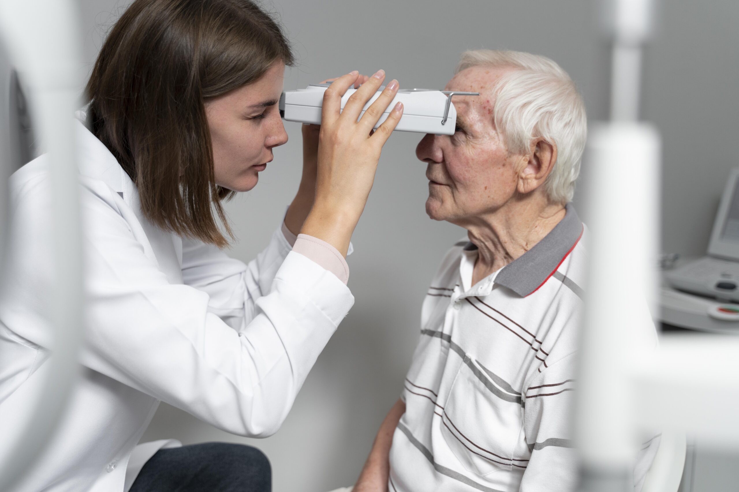
When your eye doctor peers into your pupils with that bright light, they’re seeing far more than your vision health. The tiny blood vessels in your retina are identical to those in your brain and heart, making your annual eye exam a powerful window into your stroke risk years before you’d notice any symptoms.
Your Retina Reveals Everything
Your retina contains the only blood vessels in your body that can be directly observed without surgery. These delicate vessels mirror the health of blood vessels throughout your cardiovascular system, including those feeding your brain.
When cardiovascular disease begins developing, retinal blood vessels show specific changes: narrowing, irregularities in wall thickness, and abnormal branching patterns. Advanced imaging can detect arteriovenous nicking, where arteries compress veins at crossing points, indicating elevated blood pressure damage.
Optical coherence tomography and fundus photography can measure vessel diameter changes as small as 5 micrometers. These microscopic alterations correlate directly with stroke risk, often appearing years before traditional risk factors like chest pain or shortness of breath become noticeable.
Why Age Multiplies Risk
After age 40, your risk of retinal vascular changes increases dramatically due to natural aging of blood vessel walls. Decades of exposure to blood pressure fluctuations, even within normal ranges, create cumulative damage that becomes visible in retinal imaging.
Women face additional risk during and after menopause when declining estrogen affects blood vessel flexibility. Men typically show earlier onset of retinal changes, often beginning in their late 30s, correlating with higher baseline cardiovascular disease rates.
By age 55, nearly 60% of adults show some degree of retinal vascular abnormality, even when feeling completely healthy. This age group often has multiple risk factors converging simultaneously: declining metabolism, increased inflammation, and reduced vascular repair mechanisms.
Retinal Red Flags
- Retinal hemorrhages or microbleeds visible during eye exam
- Arteriovenous nicking where blood vessels cross in the retina
- Cotton wool spots indicating nerve fiber damage
- Hard exudates showing protein and lipid deposits
- Flame-shaped hemorrhages along nerve fibers
Protect Your Vessels Daily
Mediterranean-style eating patterns rich in omega-3 fatty acids directly improve retinal blood vessel health within 6-8 weeks. Studies show that consuming fish twice weekly and daily nuts can measurably improve vessel diameter and reduce inflammation markers.
Regular aerobic exercise increases nitric oxide production, which keeps retinal vessels flexible and responsive. Even 30 minutes of brisk walking five days weekly can reverse early-stage retinal vessel narrowing in adults over 45.
Blood pressure management becomes critical, as even mildly elevated readings (130-139 systolic) cause progressive retinal damage. Home monitoring helps catch the subtle fluctuations that office visits often miss, allowing for earlier intervention before permanent vessel changes occur.
Your Prevention Checklist
- Schedule comprehensive dilated eye exam with retinal imaging annually after age 40
- Request specific retinal photography to establish baseline vessel measurements
- Monitor blood pressure at home weekly, tracking patterns over time
- Adopt Mediterranean diet emphasizing fish, nuts, olive oil, and leafy greens
- Maintain consistent sleep schedule of 7-8 hours to support vascular repair
Sleep’s Hidden Connection
Sleep quality directly impacts retinal blood vessel health through its effect on blood pressure regulation and inflammatory processes. Poor sleep causes blood pressure spikes during REM cycles, creating repetitive stress on delicate retinal vessels.
Sleep apnea, affecting 30% of adults over 50, creates oxygen fluctuations that damage retinal vessels faster than any other cardiovascular risk factor. Many people remain undiagnosed because daytime fatigue feels normal with aging.
Even one night of poor sleep triggers inflammatory markers that appear in retinal imaging within 24 hours. Consistent 7-8 hour sleep patterns allow vessels to repair overnight damage and maintain the flexibility needed to prevent stroke-related complications.
Bottom Line
Your annual eye exam is actually a cardiovascular screening that can predict stroke risk years before symptoms appear. The retinal blood vessels visible during routine eye exams mirror the health of vessels in your brain and heart, making this simple screening one of your most powerful preventive tools.
Always consult a qualified healthcare provider before making changes to your health routine.
Sources
- Retinal Vascular Caliber and Risk of Stroke — JAMA
- Retinal Microvascular Abnormalities and Incident Stroke — Lancet
- Fundoscopic Findings and Stroke Risk Assessment — BMJ