



How advanced imaging is changing what doctors can detect—and treat—before it progresses
KEY STATISTICS
- Advanced MRI imaging can detect cellular changes up to 2 years earlier than conventional methods in some cancers
- Heart patients undergoing advanced MRI imaging show 30% more accurate diagnosis of myocardial tissue damage
- Utah’s Intermountain Medical Center is the first in the state to deploy this generation of MRI technology for dual cancer and cardiac applications
If you’re 35 or older, the quality of imaging your doctor uses matters more than you might think. Most of us assume that an MRI is an MRI—a machine spins, you get pictures, you get answers. But the technology behind those pictures has evolved dramatically.
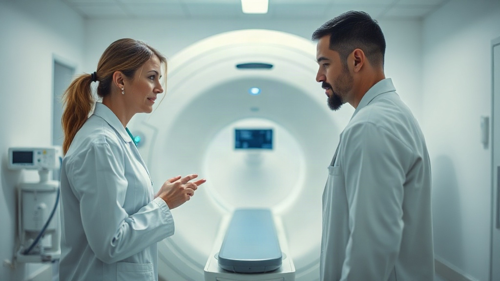
Intermountain Health’s recent introduction of innovative MRI technology at Intermountain Medical Center marks a shift in how Utah patients with cancer or heart disease get diagnosed and monitored. This isn’t just incremental improvement; it’s a fundamental change in what doctors can see, and how early they can see it.
How advanced MRI works
Modern MRI relies on magnetic fields and radio waves to create detailed images of soft tissue—the very place where cancer and heart disease hide. Newer generations of this technology increase resolution, speed, and the variety of information captured in a single scan.
- High-field MRI magnets (typically 3 Tesla or higher) generate stronger signals, producing clearer images of tumors and tissue changes at smaller scales
- Functional imaging sequences measure blood flow and metabolic activity, revealing how cancer cells or damaged heart tissue behave—not just where they are
- Reduced scan times mean less patient discomfort and fewer motion artifacts that blur images, improving diagnostic accuracy
Why 35+ matters most
Adults over 35 carry cumulative lifetime risk for both cancer and cardiovascular disease. Early detection with advanced imaging directly impacts survival and quality of life, making access to state-of-the-art diagnostic tools particularly valuable for this age group.
- Cancer incidence rises sharply after age 35, with most diagnoses occurring in the 50-70 range; earlier imaging catch prevents late-stage disease
- Heart disease risk accelerates in the 40s and 50s; advanced MRI detects scar tissue and irregular blood flow before symptoms occur
- Patients over 35 often have established baseline health data, making advanced imaging comparisons over time more meaningful and actionable
When to seek imaging
- Persistent chest discomfort or shortness of breath during routine activity—report to your doctor even if it seems mild
- Unexplained weight loss, fatigue, or changes in appetite lasting more than two weeks
- New or changing skin lesions, lumps, or areas of thickened tissue that don’t resolve
- Irregular heartbeat, palpitations, or sensation of heart skipping beats
- Swelling in the legs, ankles, or abdomen without clear cause
Habits that reduce risk
Advanced imaging alone doesn’t prevent disease—it detects what’s already forming. Complementary lifestyle changes reduce your risk profile and can slow progression of existing conditions. Think of advanced MRI as the checkpoint; your daily habits are the guardrails.
- Aerobic exercise 150 minutes weekly strengthens heart muscle and improves blood vessel function, reducing both cardiac and cancer risk
- Anti-inflammatory diet rich in vegetables, omega-3 fish, and whole grains supports cellular health and may slow tumor progression
- Stress management through meditation, sleep optimization, or counseling reduces inflammation markers linked to both cancer and heart disease
Your next steps
- Ask your primary care doctor if you meet screening criteria for cancer or cardiac MRI given your age, family history, and current symptoms
- If you have a family history of cancer or early heart disease, request advanced imaging rather than standard screening to establish a baseline
- Schedule regular check-ups to build a long-term imaging record; repeat scans at intervals help doctors track subtle changes over time
- Discuss with your cardiologist or oncologist whether functional MRI sequences would add diagnostic value to your specific condition
- Request imaging centers that use the latest MRI platforms; your location now includes one of Utah’s most advanced facilities
Speed and comfort count
The speed of a scan matters more than many patients realize. Newer MRI machines complete detailed scans in 20-30 minutes instead of 45-60, reducing motion artifact and the anxiety some patients feel in enclosed spaces. For cancer patients undergoing serial imaging to monitor treatment response, faster scans mean more frequent, safer follow-up monitoring.
- Shorter scan times reduce patient movement and breathing artifacts, improving image quality and diagnostic reliability
- Less time in the MRI bore decreases claustrophobia and anxiety, improving compliance with recommended screening schedules
- Faster protocols allow doctors to perform multiple imaging sequences in one visit, capturing more comprehensive data without extending appointment length
Bottom Line
Advanced MRI technology represents a tangible upgrade in how your doctors can detect and monitor cancer and heart disease. If you’re in Utah and have risk factors for either condition, access to this newer generation of imaging at Intermountain Medical Center is now available—making this an ideal moment to discuss your screening strategy with your primary care provider. Combined with consistent lifestyle choices and regular follow-up, early detection gives you the most control over your health outcomes in the years ahead.
Health Of State — always consult a qualified healthcare provider before making changes to your health routine.
Sources
- First in Utah: Intermountain Health Brings Innovative MRI Technology to Intermountain Medical Center — Google News — Heart Health
- General guidance on cancer screening and prevention — CDC
- Heart disease risk factors and cardiac imaging — Mayo Clinic